106
Joint injuries and degenerative articular conditions present complex challenges for the human musculoskeletal system. Whether recovering from an anterior cruciate ligament tear, managing advanced osteoarthritis of the hip, or rehabilitating a rotator cuff after surgical repair, restoring pain-free movement requires a deliberate, structured plan. Passive interventions such as rest, ice, and basic splinting may alleviate acute discomfort, but they do not rebuild functional capacity. Sustained joint restoration depends on the strategic combination of clinical physical therapy and progressive, targeted exercise.
When skilled clinicians evaluate joint mechanics, they look beyond isolated points of tenderness. True recovery involves restoring normal arthrokinematics, resolving compensatory movement patterns, and conditioning supporting muscular structures to absorb mechanical loads. Combining clinical physical therapy techniques with individualized exercise creates an environment where injured tissues can adapt, remodel, and withstand daily physical demands.
The Biomechanical Foundation of Joint Rehabilitation
A biological joint is far more than two bones meeting at a hinge. It is an intricate dynamic assembly of hyaline cartilage, fibrocartilage labra or menisci, synovial fluid, capsular ligaments, and surrounding musculotendinous units. When an injury or wear occurs, the joint loses its mechanical equilibrium.
Joint stability relies on two distinct systems working together:
-
Passive Stabilizers: These non-contractile structures, including ligaments, joint capsules, and intra-articular fibrocartilage, define normal physiological boundaries of movement. Once stretched, frayed, or torn, their structural integrity decreases, predisposing the joint to micro-instability and uneven contact pressures.
-
Active Stabilizers: These skeletal muscles and tendons dynamically control joint motion. Through coordinated contractions and reflexive micro-adjustments, active stabilizers absorb kinetic energy, minimize shear forces across articular surfaces, and maintain optimal joint positioning during movement.
When passive stabilizers sustain damage, active stabilizers must compensate to protect the joint. If surrounding muscle groups lack adequate strength, endurance, or motor control, abnormal friction accumulates within the joint capsule. Over time, this imbalance accelerates cartilage wear and leads to chronic inflammation. Combining structured physical therapy with targeted exercise addresses both passive limitations and active deficits, restoring equilibrium to the joint system.
The Distinct Roles of Physical Therapy and Targeted Exercise
While physical therapy and targeted exercise share the common goal of functional restoration, they serve complementary, distinct purposes during the healing timeline.
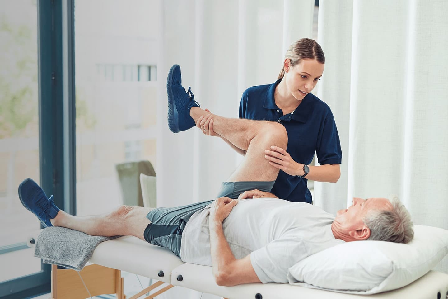
Physical therapy serves as the diagnostic and corrective phase of care. A licensed physical therapist identifies specific dysfunctions, such as restricted joint capsule mobility, soft-tissue adhesions, motor control deficits, or muscular imbalances elsewhere along the kinetic chain. Clinical physical therapy utilizes specialized modalities and manual techniques, including passive physiological mobilizations, instrument-assisted soft tissue mobilization, neuromuscular electrical stimulation, and dry needling. These hands-on interventions reduce acute swelling, calm central nervous system sensitization, and prepare tissues for active work.
Targeted exercise, on the other hand, is the active vehicle for tissue remodeling. Simply moving more is rarely sufficient; exercises must apply specific mechanical stress along precise anatomical vectors to stimulate the healing of target structures without irritating damaged tissues. While general physical activity supports cardiovascular and metabolic health, targeted exercise focuses on isolated muscle engagement, movement mechanics, and progressive resistance to rebuild functional capacity.
Without clinical physical therapy to normalize joint mechanics and release soft-tissue restrictions, targeted exercise can inadvertently reinforce compensatory movement patterns. Conversely, manual therapy without targeted exercise produces short-lived relief, as the joint lacks the muscular strength required to maintain newly acquired ranges of motion.
Phased Progression from Acute Healing to Dynamic Performance
Successful rehabilitation follows biological healing timelines. Tissues heal and adapt through distinct stages, requiring a structured progression of clinical interventions and exercise selections.
Phase One: Protection, Pain Modulation, and Passive Mobility
The earliest phase prioritizes symptom reduction, effusion control, and the preservation of resting range of motion without overloading healing tissues.
-
Manual Joint Glides: Clinicians use low-grade passive mobilizations to stimulate synovial fluid production and activate mechanoreceptors that downregulate pain pathways.
-
Isometric Muscle Activations: Low-intensity muscle contractions against fixed resistance stimulate motor unit firing while maintaining a static joint angle, preventing joint shear while fighting arthrogenic muscle inhibition.
-
Active-Assisted Range of Motion: Pulleys, slide boards, and manual clinician guidance move the limb through safe, non-provocative movement arcs to limit scar tissue tethering and prevent joint stiffness.
Phase Two: Neuromuscular Re-Education and Stability
As acute inflammatory markers recede and foundational mobility improves, the focus shifts toward neuromuscular control and dynamic joint stabilization.
-
Proprioceptive Training: Injured joint capsules lose sensory mechanoreceptors, which reduces the patient’s kinesthetic awareness. Balancing on unstable surfaces, using perturbational challenges, and performing single-leg balance drills recalibrate these sensory pathways.
-
Closed Kinetic Chain Exercises: Movements performed with the distal extremity fixed against a stable surface, such as leg presses, wall squats, or modified planks, produce axial compression forces across the joint. This loading pattern enhances congruency and stimulates co-contraction of surrounding muscle groups.
-
Eccentric Muscle Conditioning: Controlled lengthening under load strengthens tendon attachments and improves the muscular system’s capacity to absorb decelerative forces during daily movement.
Phase Three: Functional Loading and Long-Term Durability
The final phase bridges clinical rehabilitation and independent physical independence. Here, exercises mimic real-world functional tasks, athletic demands, and occupational duties.
-
Multi-Planar Strength Training: Human joints move through sagittal, frontal, and transverse planes. Exercises evolve from single-plane motions into compound, multi-directional patterns such as rotational lunges, step-downs, and overhead reaches.
-
Rate of Force Development: High-velocity movements, plyometrics, and rapid directional shifts train muscles to fire quickly, protecting the joint during unexpected stumbles or rapid sports maneuvers.
-
Work-Specific Conditioning: Reconditioning protocols are tailored to the physical demands of the patient’s job or recreation, ensuring that repaired joints can handle prolonged postures, heavy lifting, or repetitive actions.
Correcting Kinetic Chain Dysfunctions
A frequent error in joint rehabilitation is focusing exclusively on the injured anatomical site. Joints do not function in isolation; they exist within a continuous biomechanical network known as the kinetic chain. Mechanical breakdown at one link inevitably creates adverse compensation elsewhere.
Consider an individual recovering from a patellofemoral knee injury. While the knee is the site of pain, the root cause often involves poor pelvic stability and weak hip abductors, specifically the gluteus medius. When hip stabilizers fail to anchor the femur during walking or running, the thigh bone rotates inward and adducts, placing high valgus stress across the knee joint. A treatment plan that focuses only on strengthening the quadriceps while neglecting hip control leaves the patient vulnerable to ongoing knee irritation.
Similarly, chronic shoulder impingement often stems from poor thoracic spine mobility and weak scapular retractors like the middle trapezius, lower trapezius, and serratus anterior. If the thoracic spine cannot extend and the scapula cannot upwardly rotate smoothly, the subacromial space narrows, pinching the rotator cuff tendons whenever the arm elevates. By combining clinical manual releases to stiff thoracic segments with targeted exercise for scapulothoracic stabilizers, the joint can glide without mechanical impingement.
Comprehensive joint recovery requires evaluating adjacent joints both above and below the injured area, ensuring that proximal strength and distal mobility work together to protect the recovering joint.
The Critical Role of Patient Education and Self-Management
A successful joint recovery program extends beyond scheduled clinical appointments. The ultimate objective of physical therapy is to help patients safely self-manage their joint health for years to come.
Education clarifies the difference between normal adaptation discomfort and harmful structural distress. Patients recovering from joint issues often develop fear-avoidance behaviors, believing that any sensation of physical discomfort signals tissue damage. Physical therapists teach patients to identify appropriate exertion, monitor post-exercise swelling, and modify volume when joint symptoms flare.
Equally important is establishing an accessible, consistent home exercise program. Clinicians design sustainable daily routines that patients can complete with minimal equipment, such as resistance bands, light weights, and body weight. By teaching patients how to warm up stiff tissues, load active stabilizers, and stretch restrictive muscle groups, physical therapists give them the tools to maintain joint health and prevent recurrent injuries long after clinical discharge.
Frequently Asked Questions
What should an individual do if a joint begins swelling hours after completing targeted exercises?
Delayed swelling typically indicates that the mechanical volume or resistance applied during the workout exceeded the joint’s current load-bearing threshold. Patients should temporarily reduce resistance, limit deep ranges of motion, and use brief periods of limb elevation to control fluid retention. If swelling continues for more than forty-eight hours or is accompanied by sharp joint line pain, consult the treating physical therapist to adjust the rehabilitation progression.
How does a physical therapist decide when a patient is ready to advance from isometrics to heavy lifting?
The transition depends on functional criteria rather than arbitrary calendar dates. A clinician looks for the absence of resting effusion, full or near-normal passive range of motion, symmetrical limb alignment during baseline bodyweight tasks, and the ability to perform pain-free isometric contractions at maximum voluntary effort before introducing heavy dynamic loads.
Can targeted exercise cause further wear to a joint that already has mild or moderate cartilage loss?
When prescribed along appropriate anatomical angles and mechanical loads, targeted exercise does not accelerate joint deterioration. In fact, hyaline cartilage lacks a direct vascular supply and relies on cyclical, moderate compression to draw in nutrients from synovial fluid. Controlled movement promotes cartilage health, provided high-impact, shearing movements are minimized during early conditioning.
Why are open-chain exercises sometimes avoided during early-stage knee rehabilitation?
Open-chain knee extensions against external resistance near terminal extension place high shear forces across the anterior cruciate ligament and generate excessive compressive stress beneath the patella. Closed-chain alternatives, such as leg presses or supported wall slides, disperse forces across multiple joints and engage the hamstrings and quadriceps together, protecting healing tissues.
What distinguishes dry needling performed in physical therapy from traditional acupuncture?
Dry needling is grounded in Western neuroanatomy and modern musculoskeletal evaluation rather than traditional meridian concepts. A physical therapist uses thin filiform needles to target discrete, hyperirritable muscular trigger points and connective tissue bands. The goal is to release taut muscle fibers, improve local blood circulation, and interrupt local biochemical pain pathways to restore joint mobility.
How long does a patient typically need to continue their targeted joint exercises after formal therapy ends?
Targeted joint conditioning should transition into a lifelong maintenance habit rather than stopping upon clinical discharge. While the frequency and intensity can be reduced once full strength and pain-free mobility return, maintaining supporting muscle strength and joint flexibility is the most reliable way to prevent symptom recurrence and ongoing degenerative changes.
How can a person tell the difference between normal muscle soreness and abnormal joint pain during rehab?
Normal muscular adaptation, often known as delayed onset muscle soreness, presents as a dull, diffuse ache in the muscular belly that peaks twenty-four to forty-eight hours after exercise and improves with gentle movement. In contrast, problematic joint distress typically presents as a sharp, localized, or throbbing ache within the joint capsule itself, often accompanied by clicking, a sense of instability, or rapid swelling.